Bioimaging
We are developing and applying a suite of transformative new bioimaging tools exploiting quantum sensing, super-resolution optical imaging, correlated electron microscopy, cathodoluminescence, and new modalities for NMR and MRI. Applications range from noninvasive magnetic imaging of individual neurons to high-resolution NMR spectroscopy of single cells to open-access, low-magnetic-field human MRI. Some examples are given below.
Quantum Diamond Microscope (QDM)
The Quantum Diamond Microscope (QDM) provides noninvasive, wide-field magnetic imaging of 2D biological samples with diffraction-limited resolution and high sensitivity. The QDM can map static magnetic field patterns, such as populations of living magnetotactic bacteria and in magnetic nanoparticle-labeled cells and biomarkers. The QDM can also be configured for real-time magnetic imaging of currents in electrically active cells and cellular networks, such as cardiac cells and neurons with single cell resolution. These capabilities are not available with any other technology.
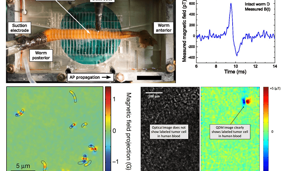
A Quantum Diamond Microscope can be used for (top row) noninvasive magnetic sensing of action potentials in live marine worms; (lower left) chains of naturally occurring magnetic nanoparticles in bacteria; (lower right) magnetic nanoparticle-labeled tumor cells in blood.
Functionalized Super-Resolution Imaging
We pioneered the use of nanoparticles to provide functionalized electron microscopy via multi-color cathodoluminescence of nanoparticles, which may allow determination of both the structure and function of brain circuits with individual synapse resolution. We also pursue new approaches to add functional capability to super-resolution optical imaging by integrating STED and RESOLFT techniques with nanoscale sensing using quantum defects in diamond.
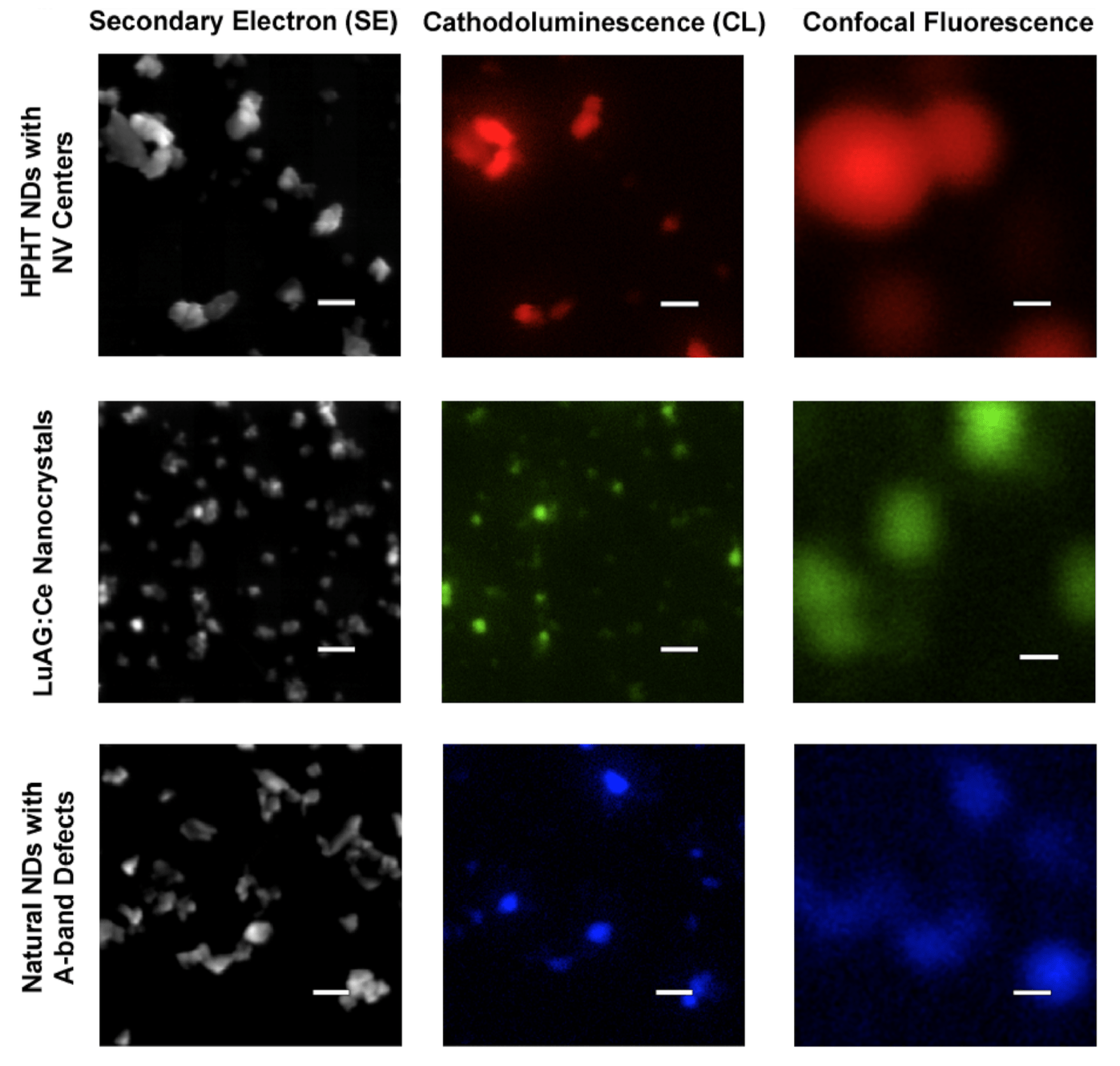
Multi-color cathodoluminescence (CL) of nanoparticles containing different types of quantum defects. The CL images are well-correlated with electron microscopy images of the nanoparticles and provide superior resolution to confocal imaging. Scale bars are 200 nm.
Publications
 link
 PDF
- Fundamental Precision Bounds for Three-Dimensional Optical Localization Microscopy with Poisson Statistics. . Physical Review Letters 121, 023904 (2018). link PDF supplement arXiv
BibTeX
- Mapping the microscale origins of magnetic resonance image contrast with subcellular diamond magnetometry. . Nature Communications 9, 131 (2018). link PDF arXiv
BibTeX
- Efficiency of Cathodoluminescence Emission by Nitrogen-Vacancy Color Centers in Nanodiamonds. . Small 13, 1700543 (2017). link PDF supplement
BibTeX
- Superresolution optical magnetic imaging and spectroscopy using individual electronic spins in diamond. . Optics Express 25, 11048–11064 (2017). link PDF arXiv
BibTeX
- Nanodiamond-enhanced MRI via in situ hyperpolarization. . Nature Communications 8, 15118 (2017). link PDF supplement
BibTeX
- Optical magnetic detection of single-neuron action potentials using quantum defects in diamond. . Proceedings of the National Academy of Sciences 113, 14133–14138 (2016). link PDF supplement arXiv
BibTeX
- Single-cell magnetic imaging using a quantum diamond microscope. . Nature Methods 12, 736–738 (2015). link PDF supplement
BibTeX
- A genetic strategy for probing the functional diversity of magnetosome formation. . PLoS genetics 11, e1004811 (2015). link PDF
BibTeX
- Atomic-Scale Nuclear Spin Imaging Using Quantum-Assisted Sensors in Diamond. . Physical Review X 5, 011001 (2015). link PDF arXiv
BibTeX
- Correlative light and electron microscopy using cathodoluminescence from nanoparticles with distinguishable colours. . Scientific Reports 2, 865 (2012). link PDF supplement
BibTeX